
Note: All items pictured on this website are in Frank W. Reiser's personal collection. Please contact if you wish to discuss exhibiting the collection at your institution or need additional information about the topic.

John Benjamin Dancer (1812—1887) owned an optician shop in Manchester, England. The business sold eyeglasses and a full line of optical equipment, such as magic lantern projectors, telescopes, sextants, and microscopes. John inherited the business from his father, Josiah Dancer (1779—1835), after having a long apprenticeship in the family business. The apprenticeship allowed him to learn the skills of both lens grinding and retail sales. Many optical instruments the company sold were of Dancer's design and manufacture. To fill out the sales inventory, he added optical devices made by other manufacturers. Dancer, an aficionado of microscopy, commercially made prepared slides sold through his optician business and that of others. He also provided accessory microscopy items to customers wishing to mount specimens themselves.

 CRYSTALS OF DINITROBENZOL
Polarized light 100x
John B. Dancer
The label for the Dinitrobenzol slide includes the chemical formula C12H4N2O8. It corresponds with the compound’s contemporary name, except American publications use the prefix “bi” for “di.” The chemical is listed as dinitrobenzene in German literature. The slide’s second label describes the chemical steps to follow for making the compound, assumedly followed by Dancer. It begins by pouring benzol into a mixture of nitric and sulfuric acids. This is dangerous to do. The resulting exothermic reaction generates enough heat to blow the acids into an airborne aerosol. An additional risk is that dinitrobenzol is highly toxic, and contact with one’s skin or inhalation can be fatal. According to Ullman’s Encyclopedia of Industrial Chemistry, dinitrobenzene is an alcohol-soluble crystalline solid used in making explosives.
Magic lantern is a Victorian moniker for a device used to show pictures on a screen or light-colored wall using the light from a kerosene lamp. As with LCD projectors today, the apparatus was used for entertainment and to provide illustrations as an accompaniment to lectures during the nineteenth century. Before the invention of the photographic process, magic lantern slides had to be drawn or painted onto glass. Children’s fairy tales, depictions of biblical events, and travel scenes were the most popular topics during Victorian times. Many well-off families owned a magic lantern for private enjoyment as well.

THE BROWNIES OPERATE A MAGIC LANTERN
Palmer Cox
Wood-Engraving 1890
A microscope at length they found;
And next, the Brownies gathered round
A stereopticon machine
That cast its rays upon a screen.
A thousand times it magnified,
Till, stretching out on every side,
An object large and larger spread,
And filled the gazing group with dread,
The locust, beetle, and the bee
Soon gained proportions strange to see,
And seemed like monsters close at hand To put an end to all the band.
Palmer Cox

![]()
John Dancer designed a series of lantern slides that conceptually crossed between magic lantern slides and prepared microscope slides. He mounted small translucent items, such as dried butterfly wings, between two circular glass plates held together by a wooden holder of a proper size to fit the projector. When the slide was placed between the magic lantern’s kerosene light source and its lens, a magnified view of the specimen would be shown on the screen. He was using the lantern slide as a microscope slide, but the magnifications he obtained were not as high as with a microscope’s lowest power. Only larger samples, such as butterflies and dragonflies, were workable within the magnification range of magic lantern optics. As one attempted to increase magnifications for smaller subjects, the image became too dim and useless. Thin leaves and flower petals worked well. Translucent insect wings were also visible, but the opaque bodies projected only as silhouettes.
Nevertheless, the impact of seeing a twelve-foot butterfly wing projected in color onto a wall thrilled Victorian audiences. Attaching a microscope to a magic lantern’s light source yielded higher magnifications, but again, the projected image was too dim to be visible. A much brighter light source than a kerosene lamp was needed if small specimens were to be viewable by an audience.
Mounting specimens in Canada balsam — the resin from the balsam fir — was another microscope slide-making technique that Dancer tried to apply to lantern slide construction. The viscous fluid is clear, water-resistant, and has a refractive index close to that of glass. Specimens mounted in balsam between a glass slide and a coverslip are optically similar to having been encased in solid glass. Dancer produced large-sized magic lantern slides of insects mounted in balsam and sealed between three-inch circular glass plates. Unfortunately, solidified balsam melts when heated. Heat build-up can happen while holding a slide on a screen for study and discussion. In time, depending on the frequency of the lantern slides’ use, Dancer’s balsam-mounted subjects began deteriorating. The examples in this collection show that the balsam has detached from the glass, crawled away from the specimen, turned brown, and congealed into gloppy balsam islands. Most likely, the cause of the problem was thinning the balsam by mixing it with a solvent, such as turpentine. Balsam is very viscous, similar to warm tar, so thinning it would make it flow faster and easier to work with. It would also facilitate the elimination of air bubbles trapped between the specimen and the glass. Dancer’s attempt at adapting microscopists’ balsam slide-mounting technique to magic lantern slide production worked poorly. In time, the smaller molecules of the thinning agent evaporated, and the balsam shrank. It took several years before the shortcomings of this balsam mounting technique became obvious. After it did, Dancer stopped using it.


John B. Dancer’s Mahogany Lantern Slide
Balsam Mounted Butterfly
The circular glass center of a full-sized (4 1/2 x 7 inches) mahogany magic lantern slide holds a pressed and dried butterfly. The insect’s wings are translucent and will project onto a screen in color, but the butterfly’s body, being opaque, will only project as a silhouette. Interference colors created by wing-scale topography will not be seen on the screen when using transmitted light.
Mounting large insects in Canada balsam is difficult because of the mounting medium’s high viscosity. Mixing the balsam with a turpentine-like solvent reduces its thickness and improves workability. Unfortunately, over time, the solvent is driven off by the light source generated by a magic lantern, which will cause the balsam to shrink and discolor. J. D. Dancer’s name and location are pressed into the lantern slide’s wood frame.
In the late eighteenth and early nineteenth centuries, sunlight was the brightest natural illumination available. Magic lanterns that could utilize sunlight were called “solar projectors,” and low-powered microscopes using the same source, “solar microscopes.” A movable mirror directed sunlight toward the projection device to provide the light needed. Sun-powered projection required an operator’s constant attention to adjust the mirror to follow the sun as it moved across the sky. It needed to be daytime, so the projection room had to be darkened and separated from the solar microscope to prevent stray light from contaminating the viewing room.
A solar microscope projected the dimmest image and had to be separated from the viewing room by a wall with only the microscope’s projection lens (eyepiece) poking through an opening. An additional peephole allowed the operator to check the screen to focus and align the picture. The operator changed slides, announced what the audience was seeing, and continually adjusted the sunlight-reflecting mirror. The first solar microscopes were developed around 1750 and were primarily used as a fee-generating public attraction.
John Dancer’s father, Josiah, built a solar microscope and charged the public for viewing sessions to supplement the income from his optician business. During his adolescent years, John operated the device for the performances. The most popular shows were of living microscopic pond life. Supersized mosquito larvae, aquatic beetles, worms, and algae swimming about the screen mesmerized audiences — until the unfortunate critters were cooked to death by the sunbeams illuminating them. Another responsibility for the young Dancer was to collect the supply of pond water needed for the day’s performances. As he operated the solar microscope, John had to continually watch the screen and replenish the solar microscope’s live-well with new critters as the concentrated sunlight overheated the older ones, as they began turning belly-up.
The oxyhydrogen limelight, developed during the 1830s, was the brightest human-made lighting source before the invention of the carbon arc. The device required a continuous flow of hydrogen and oxygen gases through two blowpipes to aim the small but intense flame at a small block of lime (calcium carbonate). An operator’s constant attention was needed to keep the gases flowing evenly. When the limelight was used to power a magic lantern, the image could fill a large screen. This advancement freed the device from small room viewing to use in large theaters.
When attached to a microscope, the limelight was bright enough to project highly magnified images of microscope slides onto a screen. The first use of such a combination reportedly occurred in August of 1843. According to the London Times, the creator of one such device demonstrated it in a private showing to Queen Victoria’s husband, Prince Albert. The prince was a microscope aficionado and cared for the microscopy collection that Queen Victoria inherited from her uncle, King George. The Times reported that the prince was very impressed with the instrument’s performance (see p. image xx).
When Louis Daguerre (1787–1852) made his photographic process public in 1839, Dancer took an immediate interest in it. The pictures, called daguerreotypes, had images formed by chemically depositing metallic silver onto a polished copper plate. To be seen as a positive image, daguerreotypes need to be viewed by carefully angled reflected light. Consequently, Daguerre’s process was unusable for creating magic lantern projection slides. In 1851, when Frederick Scott Archer (1813–1857) published his improved photographic technique, positive prints on clear glass became possible. Archer’s process was initially called wet-collodion photography because the photosensitive chemical emulsion needed to remain wet during its exposure to light. Later, its name was shortened to just “wet-plate photography.” After chemical development, the image was, as with a daguerreotype, a negative one. It would then be reversed to create a positive image by placing it against another sensitized plate, followed by another light exposure, a step called contact printing. The chemical development of the second emulsion-coated glass plate would then be a positive image and usable for projection by magic lanterns. The process could be repeated with the original negative many times to produce copies for commercial sales. John Dancer modified Archer’s method by using glass plates of a size that fit the magic lanterns his company sold. John Dancer is acknowledged as the first person to have produced photographic magic lantern slides (Allen 1976).
Magic lantern slides differ from microscope slides, mainly in size, which gave Dancer the novel idea of reversing a microscope and using it as a camera to make a very tiny picture of a large object. To get this to work, Dancer coated a glass microscope slide with Archer’s light-sensitive collodion emulsion. Doing so turned the microscope slide into a small photographic wet plate. When Dancer placed it under a microscope’s objective while pointing the eyepiece at a subject, the microscope’s objective focused a tiny picture onto what is now a wet-plate microscope slide. After chemically processing the slide, a small image was formed on the slide — a photograph so small a microscope was needed to see it.
John Dancer called the miniature photos “micro pictures.” To mass-produce them, he worked with a microscope tilted horizontally with its eyepiece protruding through a small opening in a light baffle, keeping the rest of the instrument in the dark. Dancer aimed the eyepiece at a rear-illuminated glass negative that was about four feet from the microscope. He always used negatives of his own making, although they may have been of another’s artwork. Dancer never published how he altered a microscope’s design to do this. Examining published diagrams of the equipment used by his contemporaries who were attempting to do the same thing gives a relatively clear idea about what was involved. One crucial adaptation was not to place the sensitized microscope slide on the microscope’s stage, where a slide is usually put for examination. Instead, the microscope’s sub-stage condenser was removed from its focusing mechanism, and the coated wet-plate slide was attached to its rack-and-pinion focusing mechanism. This enabled focusing to be done from behind the microscope by moving only the wet-plate slide. The optical components of the microscope remained motionless. Were Dancer to focus the subject’s image onto the wet plate using the microscope’s focusing knob, it would move the microscope’s entire optical tube. Doing so would change the distance between the eyepiece and the subject and, at the same time, change the distance between the objective and the wet plate. This would have the frustrating results of moving the focal point while simultaneously changing the size of the image being projected onto the wet plate.
The negative Dancer intended to copy was placed close to the microscope’s eyepiece and lit evenly from behind. A frosted (ground glass) microscope slide was placed on the sub-stage condenser mechanism and used to focus the apparatus. The frosted slide was then replaced with another that had a wet-collodion coating and was exposed to the image. After chemically developing the slide, it was allowed to dry, and the excess emulsion was carefully scraped away with a sharp knife. After drying, the picture remaining on the glass slide was sealed beneath a coverslip with balsam. Dancer made hundreds of microphotographs and sold them through his optician business, magazine ads, and a mail-order catalog. The microphotographs required approximately one hundred times magnification for viewing.
Dancer’s microphotographs sold successfully as novelty slides. He never intended them to be of scientific value. Nevertheless, the techniques he developed provided the groundwork as a way for copying and storing written media as microfilms and, more infamously, for smuggling stolen documents. Dancer’s method for photographically reducing printed matter found an almost immediate practical application during the Franco-Prussian War of 1870. During the conflict, the German army moved into France, encircled Paris, and isolated it from the rest of France. René Dragon (1813–1900) had legally acquired rights to use Dancer’s process several years earlier and had his optician business in Paris. Dragon was using the method to make novelty “peephole” jewelry by attaching one of his “micro pictures” to the back of a Stanhope lens (see p. xxx). During the siege of Paris, Dragon used Dancer’s process to create small, lightweight messages that could be carried by pigeons. Combining Dancer’s microphotography process with homing pigeon talents enabled Paris to communicate with the rest of France during the four-and-a-half months of the conflict. For this reason, John Dancer is credited as being the inventor of microfilm.

Stanhope Jewelry, ca. 1920
The cross is cast from base metal and studded with glass baguettes. It is intended to be used as a necklace. It has a centrally mounted Stanhope lens, which, when brought up to one’s eye, reveals a microphotograph of the Lord’s Prayer. The prayer is glued to the Stanhope’s flat rear surface. Stanhope lenses are still used for inexpensive jewelry, toys, novelties, and souvenirs.
However, John Dancer had to fight for it and do it publicly in print to retain his rightful honor. The history of the conflict begins with Dancer’s friendship with fellow Manchester resident William Sturgeon (1783–1850). Sturgeon was a philosophy professor best noted for his pioneering work in electricity. He invented the first practical and usable electric motor. When Sturgeon died, a plaque honoring his achievements was placed at his baptismal site in the courtyard of St. Mary’s Church in the nearby suburb of Prestwich. Dancer photographed the memorial plaque and used that negative to make his first microphotograph. The little picture was sharp enough so that the writing on Sturgeon’s memorial could be easily read using a microscope magnifying about sixty times.

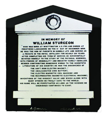 MICROPHOTOGRAPH OF STURGEON’S TABLET
John B. Dancer
Sturgeon’s Tablet is the first photographic subject used by Dancer while working on his microphotography techniques. Sturgeon’s Tablet is displayed in the garden surrounding the Church of St. Mary’s in Kirby Lonsdale, England. It is the town of William Sturgeon’s birth and the church of his christening. Sturgeon’s grave is in Prestwich, a suburb of Manchester, where he resided during his adult life. The commemorative plaque, installed after his death, commemorates his contributions to society.
George Shadbolt (1817–1901) was a London photographer, writer, and editor of the Liverpool and Manchester Photographic Journal (later becoming the British Journal of Photography) and a founding member of the Royal Photographic Society. He was also prominent as a microscopist, serving for a time as the president of the Microscopical Society of London. Shadbolt, working with the engineer Frances H. Wenham (1824–1908), developed the first dedicated dark-field sub-stage condenser in 1885 (see p. xx). He also devised a turntable used for ringing microscope coverslips, which became a popular slide-making accessory. Shadbolt was established as a prominent photographer and microscopist.
SHADBOLT’S TURNTABLE

The turntable designed by George Shadbolt was considered indispensable for performing any type of circular work on a microscope slide, but primarily the ringing of coverslips for one. Ringing sealed a coverslip’s edges, preventing dirt, humidity, and oxygen from affecting the slide’s specimen. Even when specimens were mounted in Canada balsam, the slides were “ringed.” Balsam creates an impervious seal with the coverslip, but ringing has yet to prove it improves durability. The ringing process was carried out by centering a slide on the Shadbolt turntable. A brush saturated with ringing material, such as lacquer or varnish, would then be used to apply the liquid sealant to the edge of a coverslip as the slide was rotated on the turntable.
Shadbolt was experimenting with collodion wet plates and needed to accurately measure the light-sensitive emulsion’s resolving power (ability to hold fine detail). He was writing an article comparing several different chemical image-developing processes. To do this, Shadbolt came up with a way to create microphotographs by using a microscope in reverse — the same technique developed by Dancer, but unaware of the other’s work. Shadbolt published his work, along with diagrams, in the magazine under his editorship. In his article, Shadbolt claimed to be the inventor of microphotography.
Fortunately, Dancer had shared his early microphotography work with colleagues at the Manchester Photographic Society. Many club members eagerly testified to having seen Dancer’s early microphotographs at a time that predated Shadbolt’s claim for several years. Shadbolt grudgingly began yielding his claim of primacy but replied, and again by publishing in his magazine, that he had tried Dancer’s technique but could obtain only fair results. William Henry Thornthwaite (1819–1894) was a collodion wet-plate photographer, owner of a London optician shop, and also a board member of the association that published Shadbolt’s magazine. Thornthwaite told Shadbolt that he used Shadbolt’s process precisely as described in his article. To prove it, Thornthwaite gave Shadbolt a microscope slide with the microphotograph he claimed to have made. Shadbolt examined the slide and wrote in his magazine that he possessed an example of the excellent quality micro-reproduction that his methodology could produce. He stated that the picture was sharp enough to be able to read the seven hundred fifty-five letters of writing upon the picture’s subject. The microphotograph was of William Sturgeon’s commemorative tablet.
Sturgeon’s tablet was the first microphotograph Dancer worked on and one of the first microphotographs he offered for sale through his optician shop. Dancer lists the slide as number one in his 1873 catalog of 277 slides for sale. Additionally, Dancer also had the original negative made of Sturgeon’s tablet. Shadbolt, due to Thornthwaite’s deception, unknowingly tried to validate the superiority of his methodology by holding up an example created and sold by his adversary, John Dancer.
Dancer’s last letter to the editor of Shadbolt’s photography magazine closed with the paragraph:
I cannot conclude this letter without expressing my great surprise at a statement which appears in Mr. Shadbolt’s paper, the substance of which follows: —That about March 1855, Mr. Thornthwaite showed him a copy of Sturgeon’s Tablet, that Mr. T. had procured production of, in consequence of what Mr. Shadbolt had shown to him. No doubt, there has been some misrepresentation that Mr. Thornthwaite can explain in a satisfactory manner.
All of Dancer’s and Shadbolt’s communications about the issue were read into the minutes of the April 8, 1859, meeting of the Manchester Photographic Society. The club’s members voted to do this to create an official and permanent record of the correspondence between the two men. Thus, the Manchester Microscopical Society has indelibly chronicled Dancer’s primacy as the father of microfilm.
At the age of fifty-seven, Dancer lost his vision. There are conflicting accounts as to whether the cause was diabetes or glaucoma. The loss forced him to abandon his lifetime craft, but he had time to teach his daughters, Eleanore and Catherine, the art of making microphotographs. It was Dancer’s expectation that the two would be able to take over the business as his vision worsened. According to Bracegirdle and McCormick, the sisters replaced their father’s initials, J. B. D., with E. E. D. on slides of their manufacture. (Bracegirdle, p. 23) Since a few slides affixed with such labels have turned up, it strongly suggests that most of the sales after the daughters took over the business were met by supplies drawn from their father’s original stock. Microphotographs carrying labels with the sisters’ initials are historically important finds.
Eleanore and Catherine kept the microscope slide business going until the century’s end, when microphotographs lost their novel appeal. Bracegirdle and McCormick state that Eleanore and Catherine sold the negative collection their father used for making microphotographs to Richard Suter, a London manufacturer of prepared microscope slides. Bracegirdle and McCormick report that he paid the Dancer sisters fifty pounds for the lot. Examples of microphotographs carrying Suter’s label have yet to be found.

POCKET MICROSCOPE c. 1880
France
Small microscopes that are easily portable are called pocket microscopes. The pictured example came in a wooden case with cut-out supports to cradle the instrument. A compartment for holding microscope slides contained five Dancer micro photos. It is a quality instrument with compound optics. The microscope’s eyepiece is of Huygens’ design, and the objective can be unscrewed and replaced with one or another power. The instrument is focused on by drawtube movement while the microscope slide is held in place by a pair of spring clips. When viewing a slide, the instrument must be handheld and pointed toward a light source. Most likely, its sole intended use was for viewing Dancer’s microphotographs because there is no way to control illumination other than pointing the entire microscope toward a light source. The boxed set does not carry a manufacturer’s mark other than that it has been made in France. The example came with five J. B. Dancer microphotographs.


QUEEN VICTORIA (1819-1901)
A 200× enlargement of a tiny photograph made by John Dancer in 1865
Franz Winterhalter (1805–1873) created the portrait of Queen Victoria. In 1865, John Dancer (1812–1887), recognized as the father of microfilm, turned Winterhalter’s pen-and-watercolor drawing into a miniaturized picture. The microdot in the center of the slide holds the miniature of Her Majesty. Dancer produced and sold hundreds of microscope slides containing tiny pictures, which he called microphotographs, as collectible curiosities. The slides were popular among microscope hobbyists for viewing during evening parlor conversations.
In the PBS series Victoria, a scene depicts Queen Victoria (Jenna Coleman) being painted by the artist Franz Xavier Winterhalter (Oliver Walker) (Season 2, Episode
How to footnote this page: Reiser, Frank W. (2022, October) John Benjamin Dancer. Searching an Invisible World for Its Tiniest Things. https://antiqueslides.net/John-Benjamin-Dancer/